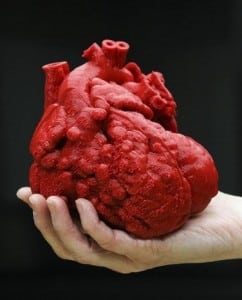
At the University of Louisiana engineering school, students were making full use of a 3-D printer in all sorts of applications but none where a life was as stake. Dr. Austin asked for and received a polymer model of Roland’s heart that showed off all the problem areas.
“Once I had a model, I knew exactly what I needed to do and how I could do it,” said Austin to the Courier-Journal. Thanks to that model, he was able to reduce the number of exploratory and the overall operating time. This would be the first successful surgery using a 3-D model of a pediatric heart patient.

The challenge with pediatric surgeries is that doctors need to delve into a very tiny and delicate organ. At this level, it is extremely difficult to see where abnormalities have occurred even with the best of scans.
“Some people think when you do heart surgery, you go in and can see everything. Well, to see everything, you have to slice through vital structures,” said Austin. “Sometimes the surgeon has to guess at what’s the best operation.”
Sadly, Roland was born with long list of heart complications including a hole in the heart, misaligned aorta and pulmonary artery. All of this prevented the heart from pumping blood, as it should.
“I didn’t think he would survive,” said Roland’s mother through a translator.
It took around 20 hours to create a duplicate of Roland’s heart that was blown up to two times its normal size. This presented a clear path for the surgical team to follow. Last week, Roland came back to the hospital for a checkup and all signs point to a happy, healthy future.
“I couldn’t express my feelings,” said mom. “He sleeps good. He plays. He smiles a lot.” In other words, just what the doctor ordered.


